
Suite 03-06 Gleneagles Medical Centre
6 Napier Road Singapore 258499

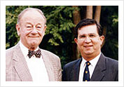 Professor Merserkinger - father of Endoscopic Sinus Surgery with Dr Stanley in Graz, Austria 1991.
 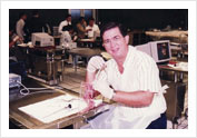 Hands-on Laboratory Endoscopic Sinus Surgery Course, Graz, Austria
 Outpatient Endoscopy - Flexible
 Diagnostic Rigid Nasoendoscopy
 Thorough evaluation of CT Scans are essential
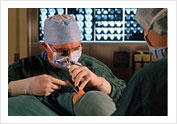 Endoscopic Sinus Surgery
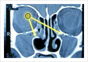 Blocked Osteo-Meatal Complex Before Surgical Treatment of Chronic Sinusitis
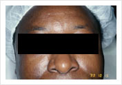 Before operation
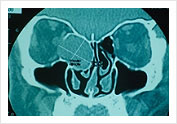 Scan before operation Mucocele in Ethmoid
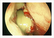 Endoscophic Drainage of Ethmoidal Mucocele during and end of operation
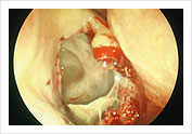 Endoscophic Drainage of Ethmoidal Mucocele during and end of operation
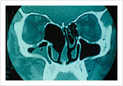 CT Scan after operation
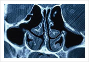 Post-operative FESS cavity on CT Scan
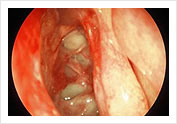 Endoscopic View of Ethmoidal Cavity after FESS operation
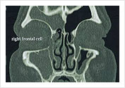 Frontal Cell causing Frontal Sinusitis – Suitable for Balloon Sinuplasty
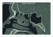 Frontal Sinusutus – Suitable for Balloon Sinusplasty from Obstructing Frontal Cell
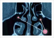
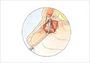
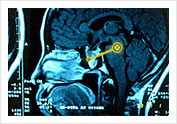 Pituitary tumour eroding the sphenoid sinus
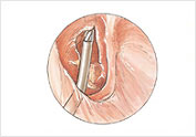
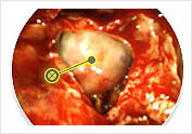 Intra-operative Transphenoidal view of tumor
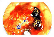 Post-operative view in the clinic 1 week after surgery
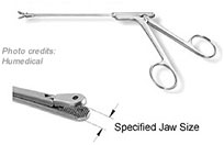 Grasping Forcepes
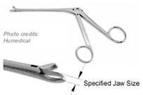 Through Cutting Forcepes
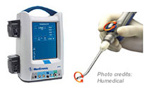
 Tri-Cut Blades
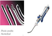
 Frontal Recess Instrument – Micro Debride
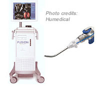 Fusion ENT Navigation System with EM Tracking Blade